Innovative dental scan helps providers find root of problem
Carol Hoover went to doctors in her home state of Louisiana in search of answers to her daily headaches, then pain on the left side of her jaw that wouldn’t go away.
She’d been wearing a mouth guard for several years because of nightly teeth grinding. “I was clenching my teeth all night,” Hoover said. “I thought I might have a sinus infection, and I went to two ENTs.”
She was referred to one dentist, who referred her to another. “He put TMD (temporomandibular joint disorder) on a physical therapy order for me, but he wanted to make sure nothing else was going on that he couldn’t see,” Hoover said.
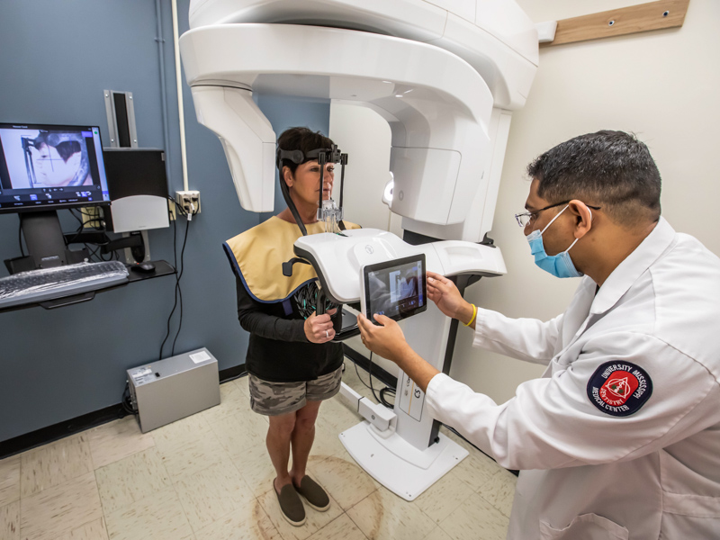
That dentist sent her to the School of Dentistry at the University of Mississippi Medical Center, which houses specialists found nowhere else in the state. On April 19, Hoover became one of the first patients in the SOD’s clinics to undergo a dental cone beam CT, which produces three-dimensional images of teeth, soft tissues, nerve pathways and bones in a single scan.
Cone beam CT is especially used when regular dental or facial X-rays aren’t sufficient to diagnose diseases of the jaw, bony structures of the face, nasal cavity and sinuses. Not only does cone beam CT provide more detailed information, it uses 10 times less radiation than a traditional CT.
“It will detect cancers, benign tumors, failures in root canals or implants and other pathologies related to diseases of the teeth,” said Dr. Rohan Jagtap, an assistant professor in the Department of Care Planning and Restorative Sciences and director of the Division of Oral and Maxillofacial Radiology.
Jagtap is the state’s sole oral-maxillofacial radiologist and the only provider in Mississippi licensed to read cone beam CT scans.
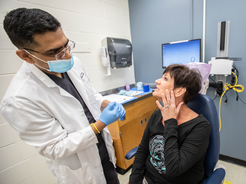
In Hoover’s case, Jagtap and his team used cone beam CT in part to confirm a diagnosis of TMD, which is caused by trauma, an improper bite, arthritis or wear and tear to the joint connecting the jawbone to the skull. It can cause jaw tenderness and locking, headaches, earaches, facial pain and trouble chewing.
Equally important, however, is using the cone beam CT to rule out bigger issues that might be causing Hoover’s pain and discomfort. “Any type of constant pain ... It’s disheartening,” said Hoover, the chief financial officer at Rogers Manufacturing in West Monroe, La. “It hurts to open your mouth, even when you’re talking. I’m a talker and a smiler, so that’s all affected.
“I wanted to make sure nothing else was wrong, and it made me happy when Dr. Jagtap said the cone beam CT would look at the whole thing. It would at least give me answers.”
Her cone beam CT took place in a room specially outfitted for the equipment, a square-shaped machine with an arm that rotates around a patient’s head in a complete 360-degree circle. As it rotates, it captures multiple images at different intervals that are reconstructed to create single 3D images.
The good news in Hoover’s case is that the cone beam CT showed no other serious issues that could be causing her jaw pain and headaches. “I’ll diagnose it, tell her dentist, and he will develop a treatment plan. I’ll send him the radiological report, and we can talk about the findings and how we can move forward with her care,” Jagtap said.
It takes about 30 minutes for the cone beam CT scans to download to a desktop computer. “You can see every tooth, every crown, sinuses, the naval cavity, all of the bones, the patient’s airway, the cervical spine,” he said of the 3D panoramic scan of Hoover’s skull. “She is having problems with her left TMJ, unfortunately.”
The cone beam CT is just one example of the technology and expertise at the SOD when it comes to using imaging to best treat a patient.
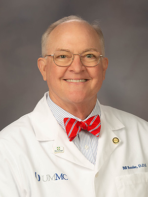
“If you are looking at this as a two-dimensional image, you can miss a lot of things. There are things you just can’t see,” said Dr. Bill Boteler, an associate professor in Care Planning and Restorative Sciences.
“It’s made diagnosis so much more accurate, easier and with less errors,” Boteler said. “Dr. Jagtap brought us not just the cone beam CT imaging, but a state-of-the-art machine. You can have a large scale of view and see the whole skull, or narrow it down, with less radiation, to focus on the area of the mouth or teeth that you want to treat.”
Dentists, oral surgeons, endodontists and orthodontists in Mississippi and surrounding states are sending patients to Jagtap, or separately sending patient scans, “when they’re not sure what’s going on, and they need help,” Jagtap said.
“You don’t want to start a treatment plan without knowing,” Jagtap said. “Some patients will drive three or four hours to get this scan and to get an expert opinion. That tells you a lot.”
The SOD’s dentists and dental specialists can immediately refer their existing clinic patients to Jagtap if a cone beam CT is needed to pinpoint a diagnosis or rule out certain diseases. “Every patient who needs radiology can do it right here,” Jagtap said. “We have an in-house service for everything they will need.”
The cone beam CT also can be a boon for SOD providers who screen patients for sleep-related breathing disorders, Boteler said. “It gives a much better image of the patient’s airway,” he said.
Confirmation of her TMD, and the knowledge that no other severe problems are the culprit in her pain, gives Hoover relief. Now, she will work with her dentist to get her TMD under control and continue exercises to regain her ability to move her jaw with less pain, or no pain.
Getting back her health means regaining quality of life.
“I’d like to eat a hamburger without having to cut it into little pieces because I can’t open my mouth wide enough,” she said.
To make an appointment, call the School of Dentistry at (601) 984-6185.