Procedure eases complications related to fluid around lungs
As people live longer with more advanced stages of cancer or chronic diseases, the longer their complications must be treated.
That can include pleural effusions, an unusually large amount of fluid around a person’s lungs that can sometimes pose a diagnostic and therapeutic dilemma. A minimally invasive procedure now being performed by specialists at the University of Mississippi Medical Center aims to find out the root cause of effusions – often confirming a disease diagnosis and providing treatment to keep it from re-occurring.
“More and more people have fluid around their lungs that needs to be addressed,” said Dr. Michal Senitko, an interventional pulmonologist and assistant professor of medicine and surgery in the Division of Pulmonary, Critical Care and Sleep Medicine.
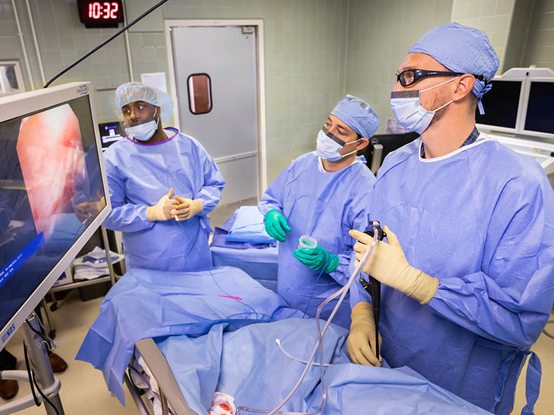
He and Dr. Trey Abraham, also an interventional pulmonologist and associate professor of pulmonary medicine, recently performed the Medical Center’s first medical thoracoscopy in 15 years. During that span, UMMC lacked a physician with specific training required to do the procedure, which is fairly rare nationally and most often performed at academic medical centers, Senitko said.
Effusion occurs when watery fluid builds up in the space between the layers of the pleura, a thin membrane that lines the surface of the lungs and the inside of the chest wall. Patients with a pleural effusion often suffer from coughing, difficulty in breathing, chest pain, fever and a feeling of chest heaviness or tightness. Sufferers sometimes have trouble lying flat or exercising, and can generally feel unwell.
A pleural effusion can be a complication of a short-term disease, such as pneumonia, or more chronic or terminal conditions, such as cancer, tuberculosis, congestive heart failure, and liver or kidney disease or failure.
When someone suffers from an effusion, a pulmonary interventionist typically drains it, giving the patient relief. “I send it for analysis, and based on that analysis, about 50 percent of the time we can get a good answer to know why the fluid buildup occurred,” Senitko said.
“But in a good percentage of the patients, the fluid comes back, and we might not know why,” he said. “We may want to look at the lining of their lungs and chest wall to get a much better understanding, and hopefully an answer, to why the fluid is coming back.”
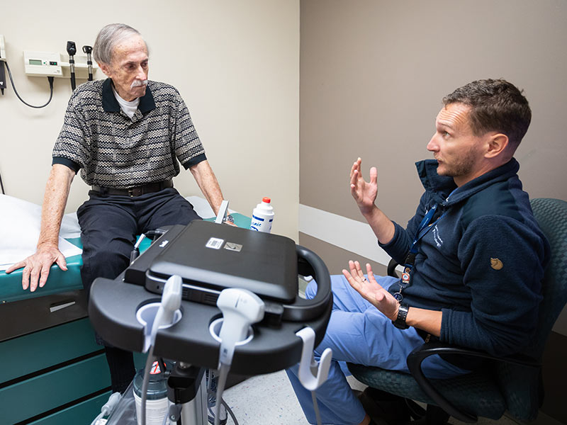
Wayne Simpson of Jackson on August 6 became Senitko’s and Abraham’s first medical thoracoscopy patient at the Medical Center. Simpson said his effusion problems were diagnosed by another UMMC physician, Dr. Lyssa Weatherly, assistant professor of geriatric medicine.
“I’d been having back problems for quite some time, and Dr. Weatherly looked at my X-rays,” he said. “It looked like my right lung was full of fluid.”
Weatherly referred Simpson to Abraham “so that they could drain off the fluid. They decided they’d put the camera down there and see what was going on,” Simpson said.
Traditionally, thoracoscopy has been performed by a surgeon using general anesthesia, and the patient is intubated and placed on mechanical ventilation. But with medical thoracoscopy, conscious sedation can be used and the patient breathes on his own.
During the 15 years that UMMC had no provider who could perform medical thoracoscopy, physicians had the option of video-assisted thoracoscopic surgery, or VATS for short. It can be a more invasive procedure and then was the only diagnostic option available to patients. Medical thoracoscopy is used mainly for patients coping with chronic conditions, Senitko said, and VATS is used more for acute conditions, or when there’s a need for more extensive work inside a patient’s chest.
“The field of interventional pulmonary medicine has grown over the last 15 years, so more pulmonologists are being trained” to perform a medical thoracoscopy, he said. “There’s a trend toward minimally invasive procedures that don’t put as much stress on the patient.”
That’s a big reason why medical thoracoscopy has made a comeback at UMMC. “Instead of doing two or three cuts or holes (for insertion of tubes and a camera), we use only one,” Senitko said. “We also noticed a higher need for complete and thorough staging (understanding the size of a cancer and how it has spread) of lung cancer patients.”
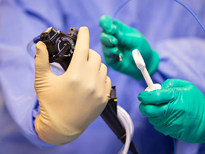
In medical thoracoscopy, an interventional pulmonologist uses ultrasound to precisely locate the presence of fluid in the pleural space. Then, the pulmonologist marks the site and makes a tiny, 1.5-centimeter incision for insertion of a small, hollow tube. Through that tube goes a scope bearing a very small camera.
“We are able to suction the fluid out and look through the camera inside the patient’s chest wall,” Senitko said. “If we see abnormalities, we biopsy it. If we feel that we need to prevent fluid from coming back, we spray a chemical or abrasive inside the patient’s chest, and it causes irritation and serves as a glue to bring the space together so that there is no more fluid produced.”
Often after the procedure, a small tube is left in place so that the patient can continue draining fluid at home for about seven days. “In 95 percent of cases, we can remove the smaller tube and there is no fluid left,” Senitko said.
In the United States, “only a small minority of pulmonologists (primarily those undergoing formal training in an interventional pulmonary fellowship) practice medical thoracoscopy,” the Journal of Thoracic Disease stated in a 2017 article. Centers in Europe and Asia have considerably more experience with the procedure, the article said.
Dr. Pierre de Delva, chief of the Division of Thoracic Surgery, “provides necessary backup for us,” Senitko said. “The cooperation between thoracic surgery and interventional pulmonary is very crucial when it comes to patients with cancer that involves part of a thoracic cavity.”
Senitko, who came to the Medical Center in early 2017, completed an interventional pulmonary fellowship at Yale University after completing a separate fellowship in pulmonary and critical care medicine at UMMC. Abraham also trained in performing the procedure both by attending subspecialty courses and by collaborating with Senitko.
There’s a bigger purpose to medical thoracoscopy, Senitko said. “This procedure is just the tip of the iceberg,” he said. “Identifying the effusions and establishing follow-up is paramount.
“In cases of benign diseases that cause pleural effusions, we need to do a better job of controlling heart and liver and kidney failure” that can lead to effusion, he said. “We need to take care of the primary problem, and to be more aggressive in managing their diseases with their liver doctor or their cardiologist.”
Regardless of the reason for a pleural effusion, Senitko said, “there’s about a 50 percent chance you will die within a year. It’s most important to have someone dedicated to seeing these patients on a regular basis.”
His procedure went “according to plan,” said Simpson, who retired from the Jackson Police Department after almost 39 years of service. “They explained every step of the way what was going on. I felt like I knew what was coming. There was no pain or discomfort.”
Dr. John Spurzem, professor and director of the Division of Pulmonary, Critical Care and Sleep Medicine, said Senitko and Abraham were recruited in part because of their training in medical thoracoscopy.
“We’re trying to avoid people having to go through the full surgery route,” Spurzem said. “This is a little lighter approach.”
One of the biggest benefit to patients, he said, is that they don’t have to undergo general anesthesia. “We expect this to become a regular procedure,” he said.
When Simpson made a follow-up visit to Senitko on August 20, the effusion had all but disappeared and Simpson’s breathing had improved. “Almost zero,” Simpson said of the effusion before Senitko removed his small drainage tube.
The procedure detected an inflammation that bears more follow-up. “We’ve achieved what we wanted to achieve,” Senitko said after performing an ultrasound that detected a tiny amount of fluid.
Simpson said he’s glad to have a better outcome, thanks to medical thoracoscopy.
“I couldn’t have asked for it to have gone any better,” said Simpson, whose effusion has markedly improved. “So far, so good.”