UMMC'S advanced PET/CT system provides clearer picture of abnormalities
An advanced PET/CT at the University of Mississippi Medical Center dramatically improves clinical, research and educational capabilities through enhanced imaging.
"It's like going from rabbit ears to high-definition TV," said Dr. Edward Green, PET/CT Center director and assistant professor of diagnostic and nuclear radiology.
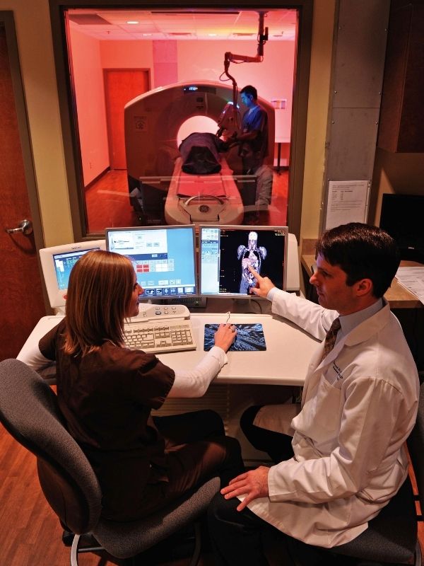
The hybrid images produced by the system - one of eight of its kind in the world - provides better clarity for diagnosing cancers and has greater applications for research in Alzheimer's disease and heart disease. For the patient, the combination of the newest PET crystal detectors, the 64-slice CT and the most advanced computer processing available enables quicker scanning. This means less time in the machine.
Green said the average time for a patient to spend in the machine is 9-15 minutes to scan from the top of the head to the middle of the thighs, compared to up to an hour in the older PET scanner, with no CT component. The PET/CT together defines the area and highlights the "bad spots" with a tiny amount of radioactive glucose, Green said.
Think of the CT (computed tomography) image as a geographic map and the PET (positron emission tomography) image as the weather system.
"Together they provide a clearer picture. With one component, it's like knowing a thunderstorm is present. With both, you can tell its exact location and its size. We can detect tumors as small as 2 to 3 mm," Green said.
Additionally, Green said the UMMC PET/CT Center is the only location in the state that offers intravenous and oral contrast during the scan, which provides a better characterization of abnormalities and helps the patient avoid the inconvenience and increased radiation exposure of a separate stand-alone CT scan with IV and oral contrast.
Dr. Ralph Vance, professor of medicine, said the new system versus the old one is like comparing daylight and dark.
"We are now seeing things we've never been able to see before," Vance said. "For us as oncologists, this is a most valuable tool in our armory of fighting cancer. It gives us new insight and a really great deal of confidence in telling patients they have no evidence of disease."
Green said that's the main reason why the PET/CT system is important.
"We're really here to significantly improve our patients' lives," he said.
The system combined with research has the potential to help advance knowledge in cardiology through stress tests that capture images of the heart and to advance knowledge in Alzheimer's disease through images of living participants in research studies.
Dr. Thomas Mosley, professor of medicine and director of the MIND Center, said he plans to use the new PET/CT system to study the pathology of Alzheimer's disease in participants. From the time the disease was first identified until today, the only way that Alzheimer's could be confirmed with 100 percent confidence has been through autopsy.
This past summer, a new radioactive dye was produced that attaches to the plaques that develop in the brains of Alzheimer's patients. Using a PET scan, the dye highlights the plaques, allowing them to be seen in participants. Mosley said he and researchers at the Medical Center have partnered with Johns Hopkins Medical Center to begin working with the new dye this spring using the sophisticated PET/CT system here at UMMC.
"We will be one of the first and largest studies to do this. It will tell us who has the pathology and who doesn't, and at what point in life it occurs. Our ability to image the plaques will allow us to better define the factors that promote the development of the disease," Mosley said.
"We are very excited about it because we believe it will have major implications for early diagnosis and development of new treatments."