Ear, Nose and Throat (ENT)
Facial Plastic and Reconstructive Surgery
Facial plastic and reconstructive surgery is an important area of treatment performed to restore or correct function to damaged tissue in the head and neck area. People may seek reconstructive surgery for a variety of reasons such as injury, disease or birth defect.
Meet the Physician
Board Certified: Facial Plastic and Reconstructive Surgery
Board Certified: Otolaryngology Head and Neck Surgery
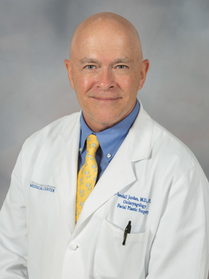
Dr. J. Randall Jordan is a Professor in the Department of Otolaryngology and Communicative Sciences at the University of Mississippi Medical Center where he serves as Medical Director and Vice Chair of Clinical Affairs. Dr. Jordan earned his M.D. at the University of Florida College of Medicine. Here, he also completed a General Surgery residency and Otolaryngology residency before completing his Facial Plastic and Reconstructive Surgery fellowship at the McCullough Aesthetic Medical Center in Birmingham, Alabama. Dr. Jordan also completed a preceptorship in Mohs Micrographic Surgery and Cutaneous Oncology at the University of Florida Department of Otolaryngology. Dr. Jordan is certified by the American Board of Otolaryngology and the American Board of Facial Plastic and Reconstructive Surgery. He specializes in facial plastic and reconstructive surgery and cutaneous oncology of the head and neck. Dr. Jordan clinical and research interests include the diagnosis and treatment of salivary gland diseases, and he leads the University of Mississippi Medical Center’s Center for Sialendoscopy and Salivary Gland Disorders.
Common Conditions and Treatments
(Follow the links below for more information on common conditions and treatments.)
Rhinoplasty - Nasal reconstruction for nasal reshaping
Refining the shape of the nose is as much an art as it is a science. It is also one of the most commonly performed plastic surgical procedures. Every nose is as unique as the individual, and our ultimate goal is to shape a nose that most appropriately harmonizes with the other facial features. We attempt to avoid the “operated look” or stereotyped nasal features. There are limits to what can be achieved with rhinoplasty. Improvement can be achieved in most every case, but absolute perfection is not a realistic goal. This is where computerized imaging technology allows the physician and patient to have a “meeting of the minds” and set realistic goals. Rhinoplasty is often performed both for functional as well as cosmetic purposes. The crooked, obstructed nose will often require internal as well as external correction. Nasal obstruction leads to chronic sinus problems as well as headaches. Whenever nasal obstruction is a part of the problem, your health insurance may cover part of the cost of surgery. In cases of sever sinus disease, surgery on the sinuses themselves may be necessary. This can often be done at the same time as the rhinoplasty.
How is the procedure performed:
The surgery is usually performed as an outpatient using general anesthesia. Surgery typically takes 2 -4 hours. A commonly asked question is whether the nasal bones are broken during the surgery. We use delicate instruments to actually incise the nasal bone and cartilage so that precise manipulation can be carried out. No nasal packing is usually required, but a small dressing is applied to collect any drainage. A small, skin-tone dressing is applied to the outside of the nose to protect the new nasal shape. This dressing will remain in place for one week. Ice cold compresses will be used to prevent swelling.
Recovery Period:
In general, the first few days after the procedure should be spent resting on the couch or in bed. After this, light activity such as desk work or light housework is acceptable for the next two weeks. As far as appearance goes, once the dressing is removed at one week, only slight and imperceptible swelling remains around the nose. Most of the swelling is gone in two weeks, but the nose continues to undergo changes during the healing process for a full year. For this reason, the tip of the nose often appears to be turned up immediately after removal of the dressing. This will come down with time. The decrease in swelling will result in a gradual refinement of the features. The nasal bones themselves do not completely heal for two months; therefore one must be careful to avoid blows to the nose or undue pressure, such as frames on heavy glasses. Supports for glasses will be provided.
Possible complications:
The most common problem after rhinoplasty is bruising and swelling. This happens to some extent in all patients but is usually minimal as long as the post-operative instructions are adhered to. Bleeding can occur in the post-operative period if too much activity is started too soon. Because there are so many variables in the outcome of rhinoplasty, it is not uncommon to need a revision surgery. This may be due to excess scar formation causing a deformity or a persistent deformity not corrected completely at the original surgery.
Septoplasty - Nasal reconstruction for nasal obstruction
Nasal obstruction is a common condition in which one is unable to breathe and pass air through the nasal passages. In most cases, this condition is due to abnormalities within the nose stemming from normal development, trauma or inflammatory changes that can lead to swelling. The most common treatment for this condition is septoplasty, the repositioning the deviated cartilage and bone in the middle of the nose.
How is the procedure performed:
A septoplasty is usually performed as an outpatient procedure taking anywhere from 60 to 90 minutes to complete. You’ll be under general anesthesia, but will be able to go home afterwards. In a typical procedure, the surgeon makes an incision on one side of your nose to access the septum. They next lift up the mucous membrane, which is the protective covering of the septum. Then the deviated portion of the septum is moved into the correct position. Any barriers, such as extra pieces of bone or cartilage, are removed. The last step is the repositioning of the mucous membrane. Dissolvable sutures are used for this along with thin sheets of soft plastic we call splints. No packing is used typically. The splints are removed at the first clinic visit at about 1 week.
Repair of Nasal Septal Perforations
A septal perforation is a hole in the septum. This can occur because of trauma, previous surgery, cautery for nosebleeds, rare types of diseases etc. Septal perforations can be bothersome because they cause nosebleeds, crusting and whistling with breathing. Septal perforations are notoriously difficult to repair. Dr Jordan has extensive successful experience with repair of septal perforations.
How is the procedure performed:
We currently use a graft of tissue from behind the ear to patch the hole. Thin sheets of plastic are placed in order to protect the graft and must stay in for 4-8 weeks. Surgery take 2-3 hours as an outpatient under anesthesia.
Melanoma and Sentinel Lymph Node Biopsy
Melanoma is a type of skin cancer that can spread quickly. When melanoma starts to spread, it often travels to a lymph node near the melanoma first. Having a sentinel lymph node biopsy (SLNB) can tell whether cancer cells have spread to a nearby lymph node. If the cancer has spread to a lymph node, you have stage III melanoma. Knowing the stage of your cancer helps your doctors create an appropriate treatment plan for you.
How is the procedure performed:
Sentinel Lymph Node Biopsy is a type of surgery that’s performed in an operating room. During this surgery, the surgeon makes a small incision and removes one or a few lymph nodes. These nodes are tested to see if they contain cancer cells.
Skin cancer and Mohs reconstruction
Mohs surgery is considered the most effective technique for treating many basal cell carcinomas (BCCs) and squamous cell carcinomas (SCCs), the two most common types of skin cancer.
How is the procedure performed?
The procedure is done in stages, all in one visit, while the patient waits between each stage. After removing a layer of tissue, the surgeon examines it under a microscope in an on-site lab. If any cancer cells remain, the surgeon knows the exact area where they are and removes another layer of tissue from that precise location, while sparing as much healthy tissue as possible. The doctor repeats this process until no cancer cells remain.
Facial Paralysis Treatments
Nerve damage due to trauma or other conditions such as Bell’s Palsy, can lead to an inability to move the muscles on one or both sides of the face. Facial paralysis can make it difficult to speak, eat, blink or even smile. In cases where the source of paralysis can be traced to a virus, medication may be used to treatment. If it is determined recovery is unlikely through medication, there are a variety of procedures available to improve facial symmetry and function.
Treatment Options:
Treatment of Facial Paralysis is very complicated and varies highly based on each individual’s situation. We perform procedures to rehabilitate the eyelids, eyebrow, nerve transfers, facial suspensions, cross face nerve grafts, gracilis free tissue transfer etc. We also do Botox injections for facial spasms and asymmetry that may follow facial nerve paralysis.
Facial Trauma and Reconstruction
Facial trauma occurs for many reasons, the most common ones being an accident, a physical altercation or cancer surgery. Trauma includes soft tissue damage to the skin or gums, bone fractures, or damage to the eyes or facial nerves. In addition to having an effect on one’s self confidence, facial trauma can cause a number of problems such as pain, difficulty breathing through the nose, trouble biting, and facial numbness and weakness to name a few.
Treatment Options:
Each facial trauma is unique, and there are many types of reconstructive surgery procedures. Your physician will help determine which one is right for you.
Recovery Period:
Recovery time depends on the level of correction needed and whether additional treatments were done at the same time. You may experience bruising and swelling of your face immediately following surgery, which may last a few weeks. Recovery time may be longer for more complex cases.
Facial Scar Revision Surgery
Facial trauma can leave behind scarring that is detrimental to one’s self-image. These defects may be improved by carefully planned and executed revision surgery. It must be understood that no scar can ever be completely eliminated, only improved. Any incision or injury that penetrates the dermis will leave a scar. All wounds undergo a healing process that takes nine to twelve months. Initially, the scar is raised and red but as it matures, it flattens, softens and becomes white. Some scars, however, do not flatten and may even widen. These are known as hypertrophic scars and keloids. Formation of this type of scar is an individual tendency and can be difficult to predict and treat. Scars that may benefit from corrective surgery are those that are lumpy or wide, pigmented, elevated or cross natural skin creases. In addition, those that alter natural landmarks such as the eyebrow or border of the lip can also be improved.
Before considering attempted improvement of a scar, several factors must be clear:
- Scars cannot be completely removed, only rearranged or improved.
- The healing process is 9–12 months and the new scar may look worse during the initial healing phases.
- The final result may be enhanced with dermabrasion or further scar revision.
Otoplasty - Treatment of prominent ears
Otoplasty is the surgical procedure used to reposition abnormally prominent ears. Prominent ears are usually an inherited trait and occur throughout the family tree. The emotional distress caused by the taunts of playmates is often unappreciated by those with normally shaped ears. For this reason, the surgery is usually per- formed at age 5, before school has begun. By this age, the ears have reached approximately 90 percent of their adult size. Often one ear is more prominent than the other, but both are usually corrected in order to obtain better symmetry. The improvement in both appearance and self-image is rewarding to patient, doctor, and family alike. Because this is a congenital deformity, some health insurance plans will help with the costs of the surgery. This will be discussed at the time of the consultation.
How is the procedure performed?
For young children, general anesthesia in a hospital is usually required, but the patient can typically go home after surgery. All incisions are made on the back of the ear so as to be well hidden. The ear is pulled back and held in place with permanent sutures beneath the skin.
Recovery period:
The head dressing will be changed and replaced the first morning after surgery. On the third day the head dress is replaced with an elastic sweatband-type dressing to be worn at night. Young children must be kept fairly quiet, i.e. away from playmates who may rough- house and disturb the operated parts. This is important for two weeks. The recovery period is usually uneventful if instructions are followed. There is virtually no pain with this procedure.
Possible complications:
Damage to the ear by a playmate is perhaps the highest risk in this procedure. The risk of recurrence of the original deformity is approximately 5 percent. This is due to the plastic nature of the cartilage of the ear. Less common complications include bleeding and infection. The latter would be signified by red- ness and tenderness of the ear.