Cardiometabolic and Imaging Core
Imaging Core
The Imaging Core is a core facility in the Department of Physiology and Biophysics that provides state-of-the-art equipment and services that are available to all UMMC researchers, collaborators and other educational and research institutions on a fee-for-service basis. The aims of this core are to provide access to high resolution imaging technologies, training, consultation, data analysis, image processing, and centralized technical expertise to support imaging needs.
Resources available include the following:
Leica TCS SP8 Laser Scanning Confocal and Multiphoton Microscope
The Leica SP8 is an advanced confocal microscope equipped for multiphoton imaging. The system provides high speed and high-resolution imaging of live or fixed cultured fluorescently labeled cells, tissue sections, or small organs. It has multiple applications. Imaging of fixed, labeled samples provides information regarding structure, protein localization, oxidative stress in 3 dimensions. Imaging of live cells and tissues provides assessment of ion fluxes, protein translocation, vascular permeability, cellular migration and blood flow.
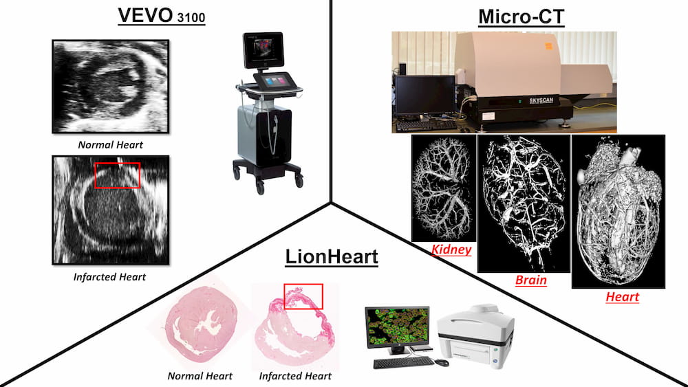
The VEVO 2100 and 3100 are advanced micro-ultrasound imaging systems that provide high quality imaging for small animal in vivo studies. They have multiple applications including assessment of kidney function, liver fibrosis and cardiac function in 2D and 4D, measurement of cardiac hemodynamics, cardiac dimensions, myocardial and vascular strain, placental structure and function, embryo injections, and tumor detection and sizing in 2D and 4D.
The Micro-CT scanner is a high-resolution low-dose X-ray scanner for in vivo and in vitro 2D and 3D reconstruction with a spatial resolution and details detectability of 9µm. It has the capability of scanning and performing 3D reconstruction in vitro of the ultrastructure and microcirculation of virtually any organ as well as whole small animals (e.g. mice, rats) in vivo.
The Lionheart FX Automated Microscope can be used to meet many imaging needs including fluorescence, brightfield, color brightfield, and phase contrast microscopy applications. It is also equipped with an environmental control cover that provides incubation to 40°C, effective containment for CO2/O2 control, and a humidity chamber for long-term live cell imaging applications.